Quantitative Digital Image Analysis Using AI-Powered Digital Pathology Software
MD Biosciences provides sophisticated digital image analysis services utilizing software programs such as Visiopharm, Aperio ImageScope, ImageJ and Adobe Photoshop to provide quantitative, semi- quantitative and qualitative data outputs from tissue specimens. Standard and custom assessments are available, including but not limited to:
Area Analysis: quantification of area parameters (fibrosis assessment, necrotic tissue percentage, inflammation, would healing, tumor area, cellularity and many more applications)
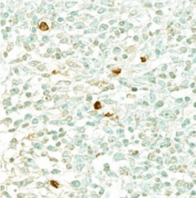
Percent Positive: the number of cells with positive staining above a defined threshold value for a given marker (or markers) of interest divided by the total number of cells in a tissue (as defined by nuclei identification)
 --->
---> 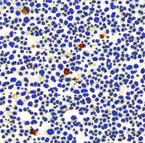
In example above, Blue=negative nuclei; Red=positive nuclei;
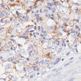
H-Scoring: Assessment of the percentage of positively stained cells and the intensity of staining for each positive cell.
 --->
---> 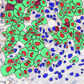
In example above, Blue=negative nuclei; Red=positive nuclei; Green=1+ staining; Purple=2+ Staining
Cellularity: Total number of cells in a given area of tissue.
Co-Localization: Analysis of two or more targets of interest within a single cell or cellular structure (membrane, cytoplasm, nuclei, ECM)
Tissue Structure Analysis: Assessment of large tissue structures such as vasculature, glomeruli, lymphoid follicles, tumor characteristics, etc.
Heat Map: Analyze the number and density and of cells across locally-defined regions of a tissue specimen to observe fluctuations or patterns.
Custom Assessments: Objective-specific feasibility assessments can be performed for complex image analysis applications to determine the optimal methods for a given analysis of interest.
All images analyzed are reviewed by our skilled scientific team and approved by qualified pathologists for appropriateness of results. Manual pathologist review and comparison assessments are also available.


CONTACT US:
+1-651-641-1770
1-888-876-3246


